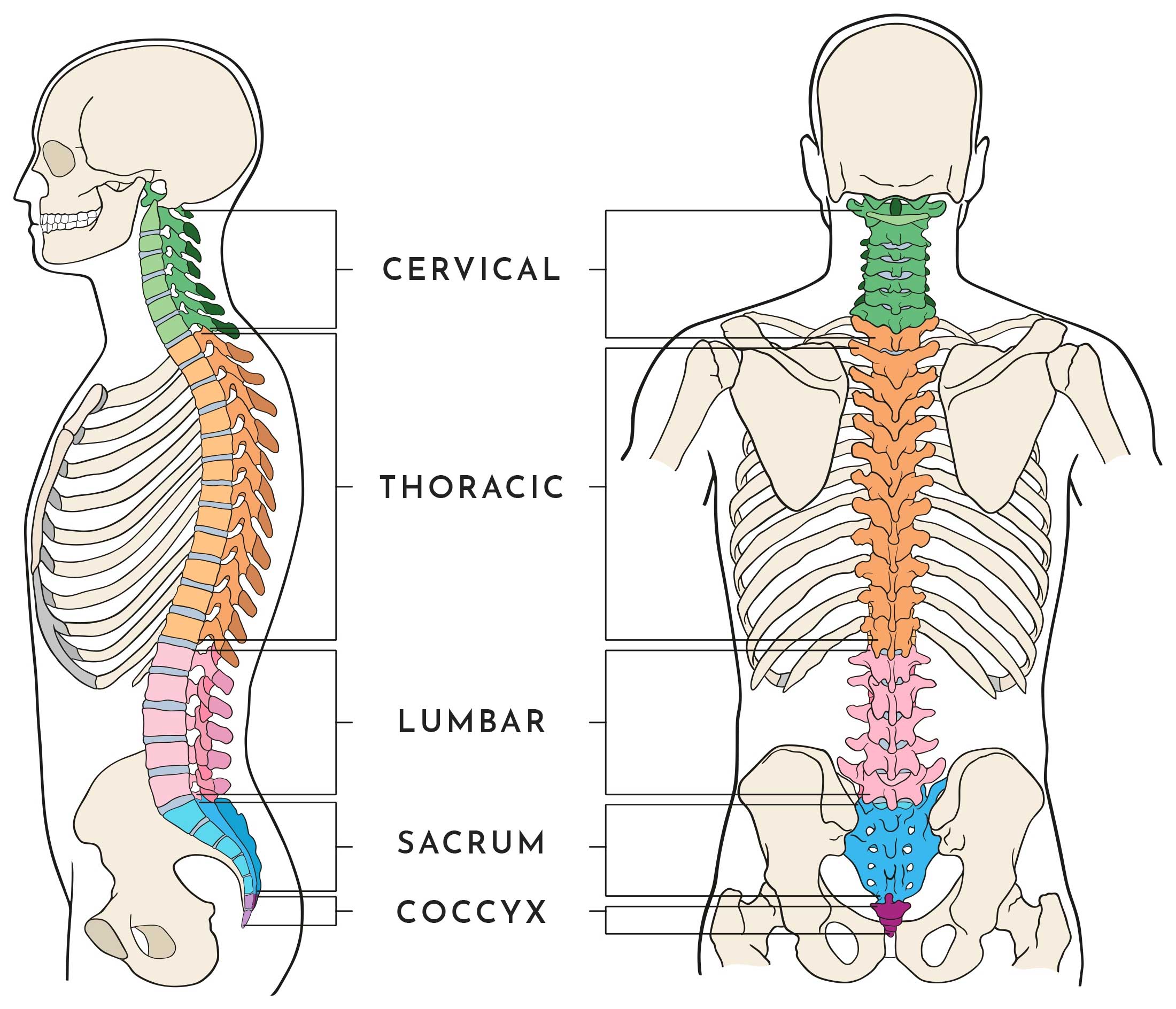
Anatomy of the Spine
The spine is made up of 33 individual bones which are all stacked one on top of the other. The spinal column allows your body to stand upright and provides the main support, allowing it to bend and twist whilst protecting the spinal cord from injury. Strong bones, muscles, nerves, tendons and ligaments surround the tissue around the spine contributing to its healthy structure.
The Spine is primarily split into 5 regions:
- Cervical
- Thoracic
- Lumbar
- Sacral
- Coccyx
The entire anatomy of the spine is then made up for eight core areas which include muscles, vertebrae, intervertebral discs, facet joints, ligaments, spinal cord, nerves, coverings and spaces.
Muscles
There are two main muscle groups that affect the spine which are the Flexor and Extensors. The Flexor muscles are in the front and include the abdominal muscles, the Extensors are attached to the back of the spine.
Vertebrae
The Vertebrae are divided into regions which include the cervical, thoracic, lumbar, sacrum, and coccyx and each region have unique features that help them perform their main functions.
Intervertebral Discs
Intervertebral Discs keep the bones from rubbing together, acting like a car tire. The outer ring (called the annulus) pulls the vertebral bones together against the elastic resistance of the gel-filled nucleus.
Facet Joints
Each vertebra has four facet joints and these joints are designed to give the back a range of motion.
Ligaments
Ligaments are are strong fibrous bands which hold the Vertebrae together to help stabilise the spine.The three major ligaments of the spine are the Ligamentum Flavum, Anterior Longitudinal Ligament (ALL), and Posterior Longitudinal Ligament.
Spinal Cord
The Spinal Cord is approximately 18 inches in length and around the thickness of your thumb. It runs from the brain to the 1st Lumber Vertebra and serves as an information super-highway, relaying messages between the brain and the body.
Spinal Nerves
There are Thirty-one pairs of Spinal Nerves which branch off of the Spinal Cord. The spinal nerves carry messages back and forth between your body and spinal cord to control sensation and movement.
Coverings & Spaces
The Spinal Cord is surrounded with membrane called Meninges (which is the same as the brain) and the space between the dura mater and the bone is the epidural space. This space is most often accessed to deliver anaesthetic numbing agents like epidural, and to inject steroid medications.
Spinal Imaging
The spine is a complex mechanical structure that surrounds and protects the spinal cord and nerve roots. Imaging of the spine is performed frequently and there are a number of ways in which it is achieved.
X-Ray
Spinal X-ray is a test that uses radiation to produce images of the bones and organs of the body. It provides detailed images of the bones and can be taken separately for the three main areas of the spine which are the Cervical, Thoracic and Lumbar.
CT
A Spinal CT scan is a type of X-ray that produces cross-sectional images of a specific part of the body. It is used to help diagnose—or rule out—spinal column damage in injured patients and is a fast, painless, noninvasive and accurate spinal imaging technique.
MRI
MRI stands for Magnetic Resonance Imaging and is a noninvasive spinal imaging technique used to diagnose medical conditions. It uses a powerful magnetic field, radio waves and a computer to produce detailed pictures of internal body structures.
SPECT
A Spinal SPECT scan is a type of nuclear imaging test, which uses a radioactive substance and a special camera to create a 3-D picture of the spinal area in question. While imaging tests such as X-rays can show what the structures inside your body look like, a SPECT scan produces images that show how your organs work.
Mr Stephen F. McGillion
BSc(Hons) MBBS MRCS(Eng) FRCS(Tr&Orth)
Consultant Orthopaedic Spinal Surgeon

